Anatomy of Complications Workshop
The Anatomy of Complications Workshop is a two day intensive, hands-on learning experience. The program is divided into three discrete but integrated and related modules:
Anatomy & Surgical Skills (Day 1)
Live Animal Surgery (Day 2)
Case Presentations (Day 2)
During the first 1.5 days participants work in trios (per cadaver) and pairs (per live sheep) that are allocated by the workshop organizers. All participants rotate through six different groupings in order to maximize interaction and to allow for the different skill and speed of participants.
Prior to attending the workshop all participants receive a login to access online learning materials, including videos demonstrating the various procedures to be performed during the practical surgical sessions. It is crucial that these materials are reviewed by participants prior to attendance at the workshop.
A week or so after the workshop each participant is required to complete an online evaluation questionnaire. This gives valuable feedback to the course organizers and allows for continued improvement in the quality and content of the workshop.
Anatomy & Surgical Skills (Day 1)
Objectives:
- To demonstrate and learn the pelvic surgical anatomy relevant to obstetric and gynaecological surgery
- To demonstrate, practice and learn the various surgical skills which may be needed to deal with unexpected intraoperative injury to bowel, bladder or ureter
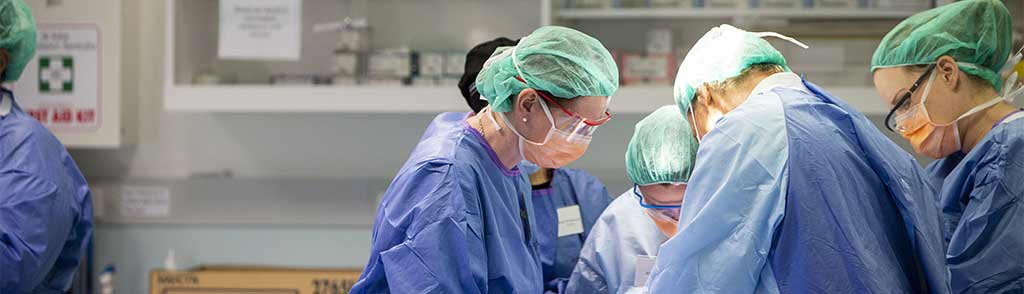
During day 1 participants will work in trios through a variety of clinical scenarios utilising fresh-frozen cadavers. Half of the time is spent dealing with the anatomy and injuries relating to the pelvis, the remainder looking at the retroperitoneum and GI tract.
The module is aimed at revising and improving the participants’ knowledge and understanding of clinically relevant anatomy. The anatomical structures which are commonly involved in surgical injury will be reviewed, in particular the major vessels, nerves, ureter and bladder. The surgical anatomy relevant to urinary incontinence surgery, hysterectomy, ovarian masses and retroperitoneal exploration of the pelvic side wall will be covered.
This session requires satisfactory completion of a number of tasks during the cadaver dissection. The various surgical procedures are carried out by all the participants under supervision by facilitators.
In the course of day 1 participants will:
- Expose pararectal and paravesical spaces
- Perform ureterolysis and pelvic peritonectomy
- If time, mobilise bladder and perform cystotomy and repair
- Perform lymphadenectomy
- Expose iliac vessels and branches
- Expose obturator nerve
- Ligate anterior division of the internal iliac artery
- Identify pelvic floor muscle and bony pelvis landmarks
- Identify and dissect along anatomical planes to mobilise colon
- Identify blood supply to the bowel
- Dissect along interior vena cava and aorta
- Vessel injury and repair
- Repair serosal tear and Veress injury to the small bowel
- Repair rectal tear
- Repair trocar injury to the stomach
- Eviscerate and close abdomen
This session lasts 8 hours and takes place in the Hill Surgical Workshop at the Clinical Training & Education Centre (CTEC) at The University of Western Australia (UWA).
Live Animal Surgery (Day 2)
Objectives:
- To demonstrate the pelvic anatomy in the ewe.
- To confidently dissect the ureter and major pelvic vessels.
- To repair injury to bowel, perform a bowel reanastamosis, repair injury to the bladder and to vessels.
- To practice the technique of internal iliac artery ligation.
- To raise a colostomy
- To control haemorrhage from a major vessel using an overlay of autologous tissue (OAT) patch (time permitting)
- To be knowledgeable about these procedures and to understand their place in the management of intraoperative injury
During this session these surgical procedures will be performed by all participants with the supervision of experienced facilitators.
The session lasts for 5 hours and is held at The Centre for Advanced Veterinary Education at Murdoch University.
Case Presentations (Day 2)
Objectives:
- To present and discuss common clinical situations occurring during and after obstetric and gynaecological surgery.
- To understand the principles of safe management of injury to bowel, bladder, ureter and major vessels.
- To understand and learn approaches which lead to a reduction in surgical injury.
This is the final session (2.5 hrs) and is held at the Veterinary Clinical Sciences Building at Murdoch University.
Prior to attendance, all participants are required to submit one case for presentation. This case usually describes a surgical or obstetric complication, or a difficult management decision. The presentation is given using Power Point or equivalent with a maximum of four slides. Each case is allocated 15 minutes to include presentation and discussion. 10 cases will be presented.
This session is facilitated by the directors, and is controlled so that the discussion is constructive and non-threatening. During this session the other participants and facilitators are asked to provide ‘micro- summaries’ of the case under consideration. At the end of each presentation and discussion the group is asked to define the ‘learning points’ from the case. The input of the invited facilitators from other specialties (urology and general surgery) is very valuable and gratefully acknowledged.
This has proved to be a most popular and valuable session; allowing for integration of all the workshop modules.